Bone grafts in implantology
Bone reconstruction for dental implants
Why perform a bone graft?
A minimum bone quantity is essential for the placement and longevity of a dental implant.
In some cases of bone deficiency, it is necessary to reconstruct the missing bone volumes.
A pre-implant bone graft (before the placement of the dental implant) or a peri-implant bone graft (at the same time as the implant placement) must then be performed.
Different types of bone reconstruction materials:
We have various types of materials and techniques available for bone reconstruction. We choose based on the surgical technique used and the preferences expressed by the patients.
Autogenous bone
We use bone from the patient themselves. For this, we need to harvest bone from a second surgical site, primarily from the jawbone. This surgical procedure is longer and has more postoperative consequences because it involves two sites.
Allogeneic bone
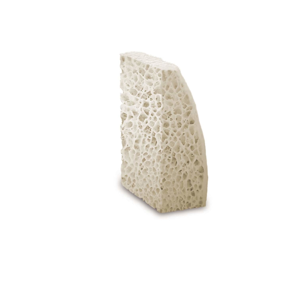
We use bone from another human. Most often, the bone comes from the femoral heads of patients. This bone is treated to eliminate any potential contaminants, but this process also causes it to lose some of its properties. The surgical procedure is shorter, but the results can be inconsistent.
Bone biomaterials
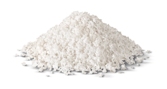
We use bone biomaterials as substitutes to replace autogenous bone grafts.
This surgical procedure is shorter, but we must adapt the placement techniques.
We use biomaterials from different origins:
Xenogeneic, of animal origin
We primarily obtain these biomaterials from cattle. We treat the bone to eliminate any risk of contamination. With over 20 years of use and numerous scientific publications, these materials are currently the materials of choice. They allow for shorter surgical procedures with reproducible results. At Névé Dental Clinic, we primarily use biomaterials from Geistlich Pharma, the brand with the most scientific publications.
Synthetic
We manufacture these biomaterials chemically. However, these materials have limited properties and inconsistent results, making their use very rare.
Les facteurs de croissance
Des petites protéines, appelées facteurs de croissance, peuvent accélérer certains processus, tels que la cicatrisation et la formation osseuse. Cependant, nous ne pouvons pas les utiliser en Europe et dans de nombreux pays en raison des réglementations en vigueur.
Certains protocoles ou biomatériaux prétendent en contenir, mais nous n’avons pas de preuve scientifique suffisante pour justifier leur utilisation à l’heure actuelle.
Alternatives to bone grafts?
In some cases, we use short or narrow dental implants to avoid bone grafts.
We have designed small-dimension implants with interesting mechanical properties thanks to recent technological advances.
However, we do not adapt them to all situations of low bone volume.
The different bone reconstruction techniques:
Sometimes, we perform bone reconstruction in height and/or thickness. Indeed, we use numerous techniques to carry out this reconstruction.
We apply suitable technical solutions to compensate for the lack of bone depending on the nature of the defect, its volume, and its location. Therefore, we always choose the most appropriate method for each case.
When we observe significant bone deficiency, we primarily perform the reconstruction procedure before the implant placement. Then, we wait for a period of 4 to 9 months to allow the bone to regenerate properly before placing the dental implant. This waiting period is crucial to ensure the implant’s stability.
On the other hand, when we observe moderate bone deficiency, we perform the reconstruction and the dental implant placement during the same intervention. This saves time and simplifies the process for the patient.
We adapt our bone reconstruction techniques to each situation. Thus, we ensure optimal results for our patients by using precise and effective methods.
-
Preservation of extraction socket
After extracting a tooth, we inevitably observe a reduction in bone volume. Consequently, this loss of volume can compromise the placement or longevity of the future dental implant. Fortunately, we use specific procedures to limit this bone resorption phenomenon.
During atraumatic extraction, we place a biomaterial in the extraction socket. Then, we protect this biomaterial with collagen or a gum graft. Thanks to this method, we maintain a bone volume favorable for the placement of a dental implant. Thus, we avoid, in most cases, complex grafts at a later date.
Clinical case
Extraction with socket preservation
-
Sinus floor élevation or Sinus lift
The sinus is a cavity in the maxillary bone located above the premolars and molars. Often, after the extraction of maxillary premolars and molars, the available bone height under the sinus is insufficient to place dental implants. To remedy this, we perform a sinus floor elevation, also known as a sinus lift or sub-sinus bone graft.
We gently lift the sinus membrane and place a biomaterial under this membrane to hold it in the desired position. This creates an ossification area, allowing your own bone cells to reform the desired bone. Typically, we achieve sufficient bone volume in 4 months.
We use two main methods for sinus floor elevation:
Lateral approach (Tatum technique)
We mainly use this method for significant bone loss. We access the bone from the side of the bone ridge. Although this technique is reliable and reproducible, we generally cannot place dental implants at the same time as bone reconstruction.
Vertical approach (Summers technique)
We mainly use this method for moderate bone loss. We access the bone on the bone ridge by drilling for the implant. This technique is less invasive, faster, and results in fewer postoperative effects. Additionally, we can often place the dental implant at the same time as the bone reconstruction.
In conclusion, sinus floor elevation is essential to ensure sufficient bone volume before placing dental implants. Whether through the lateral or vertical approach, we guarantee a solid and durable foundation for your implants.
Clinical case
Lateral approach sinus lift
-
Guided bone regeneration
We use guided bone regeneration to create a regenerative space so that your own cells can recreate bone. For this, we employ bone biomaterials, either mixed with or without autogenous bone in the form of chips. Then, we maintain them in a space created by a synthetic or collagen membrane.
Indeed, these techniques, which have been the subject of extensive research and improvements in recent years, vary depending on the case to be treated. They are certainly more delicate to perform and require an experienced surgeon. However, they produce very high-quality bone, in substantial quantity, without the need for harvesting a large volume from another site.
Furthermore, in some cases, we can place the dental implant at the same time as the guided bone regeneration. This approach allows us to optimize treatment time and reduce the number of necessary interventions.
Clinical case
Simple horizontal guided bone regeneration
Clinical case
Vertical guided bone regeneration
-
Bone grafting
We perform a bone graft by transplanting a fragment of bone.
First, we can use the patient’s own bone (autografts) or an allograft block from another human.
Then, we immobilize the bone block with screws at the site to be augmented. After about 4 months, the added bone block bonds with the jaw. At that point, we can place the implant in a sufficient bone volume.
We have been using these techniques for many years. Consequently, we have found that autografts produce good quality and quantity bone. However, they require harvesting from another site on the patient.
On the other hand, allografts do not need a second harvesting site, but the results are sometimes more inconsistent.
Clinical case
Autogenous onlay graft
